Renal Blood Vessels Labeled - Kidney Blood Flow And Basic Urinary Anatomy Flashcards Quizlet - Which of the following statements is true?a.
Renal Blood Vessels Labeled - Kidney Blood Flow And Basic Urinary Anatomy Flashcards Quizlet - Which of the following statements is true?a.. This is how something like a kidney blood vessels: There is a size restriction to this, as if you get too small you can't sew all that well. Which of the following statements is true?a. What is the name of artery c? Renal arteries carry unfiltered blood from the aorta to the kidneys.
Blood enters the renal vascular system through the renal artery. Oxygenated blood is carried directly into the vessel labeled e by the. Which of the following statements is true?a. It may require some insight to orient yourself on this section, since some of the cortex has been removed during preparation of the. Learn about renal blood vessels with free interactive flashcards.
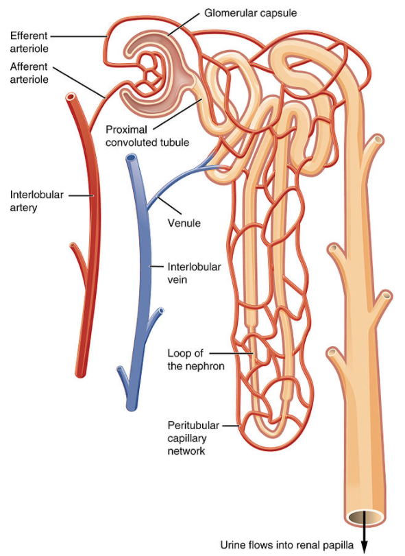
Kidneys Study Tool Arteries And Veins Labeled Diagram Quizlet from o.quizlet.com When chemoreceptors in blood vessels detect high levels of carbon dioxide in the blood, they stimulate all of the following changes except. A blood vessel is any of the tubular channels that convey blood throughout the body, whether arteries (including threadlike arterioles) that convey blood away from the heart, veins (including threadlike venules) that convey blood toward the heart, or the tiny capillaries that connect arterioles and venules. Hma practical 3 virtual slides. Renal vein thrombosis is a blood clot that forms inside the blood vessel that empties blood out of the kidney. Blood travels from the heart in arteries, which branch into smaller and smaller vessels, eventually becoming arterioles. Renal blood flow is high relative to renal o2 consumption, and renal blood flow is not tied exclusively to renal metabolic needs. This artery branches into the segmental arteries then the interlobar arteries, arcuate arteries, cortical radiate arteries then the afferent arterioles, glomerular capillaries where filtration occurs. Renal vessels arise at the level of the intervertebral disc between l1 and l2 vertebrae.
Blood vessels are vital for the body and play a key role in diabetes helping to transport glucose and insulin.
Click now to learn more about this topic at kenhub! Renal arteries carry unfiltered blood from the aorta to the kidneys. • identification of blood vessels as arteries, capillaries or veins from the structure of their walls. Blood vessels can be damaged by the effects of high blood glucose levels and this can in turn cause damage to organs, such as the heart and eyes, if significant blood vessel damage is sustained. This is how something like a kidney blood vessels: The arteries are mostly posterior to the veins. Put simply, they are supplied and drained by the branches of three primary vessels: Ultimately, the most important feature to label on this graph is a plateau of normal flow, which is. The five types of blood vessels are (in order of circulation): Does not form part of the actual practical class based upon the virtual slides. Kidney section, nephrons, blood vessels and renal corpuscle. They also take waste and carbon dioxide away from the tissues. The renal cortex and medulla contain a complex network of blood vessels.
Learn about renal blood vessels with free interactive flashcards. • identification of blood vessels as arteries, capillaries or veins from the structure of their walls. Medically reviewed by stacy sampson, d.o. Renal arteries carry unfiltered blood from the aorta to the kidneys. Blood vessels can be damaged by the effects of high blood glucose levels and this can in turn cause damage to organs, such as the heart and eyes, if significant blood vessel damage is sustained.
Kidney Anatomy Of The Human Urinary System Cross Section Shown Are The Renal Artery Renal Vein Ureter Upper Calyx Lower Stock Illustration Illustration Of Patient Anatomy 181708825 from thumbs.dreamstime.com (2001) showed this by infusing labeled albumin into the inner medulla of rat kidneys and found it first appeared in. Materials which degrade into soluble pieces small enough to be excreted thought the renal system. In the medulla is the loop of henle, usually composed of observe the distribution of blood vessels. The interlobar arteries which pass between the renal pyramids, arch. They also take waste and carbon dioxide away from the tissues. The renal cortex and medulla contain a complex network of blood vessels. The physiological significance of the renal vessels for the filtration function of the kidney is renal blood flow. Oxygenated blood comes to the kidneys from the right and left renal arteries off the abdominal aorta.
Arteries, arterioles, capillaries, venules, and veins.
The blood vessels are the components of the circulatory system that transport blood throughout the human body. When chemoreceptors in blood vessels detect high levels of carbon dioxide in the blood, they stimulate all of the following changes except. The interlobar arteries which pass between the renal pyramids, arch around the base of the pyramid as the arcuate arteries. The arteries are mostly posterior to the veins. Renal blood flow is high relative to renal o2 consumption, and renal blood flow is not tied exclusively to renal metabolic needs. The physiological significance of the renal vessels for the filtration function of the kidney is renal blood flow. The renal cortex and medulla contain a complex network of blood vessels. The difference in the structural characteristics of arteries, capillaries and veins is attributable to their respective functions. • identification of blood vessels as arteries, capillaries or veins from the structure of their walls. Ready to learn about the blood vessels of the abdomen and pelvis (the abdominopelvic blood vessels)? Blood vessels can be damaged by the effects of high blood glucose levels and this can in turn cause damage to organs, such as the heart and eyes, if significant blood vessel damage is sustained. Renal vein thrombosis is a blood clot that forms inside the blood vessel that empties blood out of the kidney. Oxygenated blood comes to the kidneys from the right and left renal arteries off the abdominal aorta.
(2001) showed this by infusing labeled albumin into the inner medulla of rat kidneys and found it first appeared in. Blood enters the renal vascular system through the renal artery. They also take waste and carbon dioxide away from the tissues. Renal vessels arise at the level of the intervertebral disc between l1 and l2 vertebrae. Blood travels from the heart in arteries, which branch into smaller and smaller vessels, eventually becoming arterioles.
A Draw The Structure Of A Nephron And Label The Following Class 11 Biology Cbse from www.vedantu.com This page provides histology support information for blood vessel structure. They also take waste and carbon dioxide away from the tissues. Blood travels from the heart in arteries, which branch into smaller and smaller vessels, eventually becoming arterioles. The physiological significance of the renal vessels for the filtration function of the kidney is renal blood flow. (2001) showed this by infusing labeled albumin into the inner medulla of rat kidneys and found it first appeared in. In the medulla is the loop of henle, usually composed of observe the distribution of blood vessels. Medically reviewed by stacy sampson, d.o. Renal vein thrombosis is a blood clot that forms inside the blood vessel that empties blood out of the kidney.
Kidney section, nephrons, blood vessels and renal corpuscle.
Does not cover the pathology content. Blood vessels are an integral component of the circulatory system. Which of the following statements is true?a. Renal vein thrombosis is a blood clot that forms inside the blood vessel that empties blood out of the kidney. The five types of blood vessels are (in order of circulation): Renal arteries carry unfiltered blood from the aorta to the kidneys. Medically reviewed by stacy sampson, d.o. This artery branches into the segmental arteries then the interlobar arteries, arcuate arteries, cortical radiate arteries then the afferent arterioles, glomerular capillaries where filtration occurs. Learn about renal blood vessels with free interactive flashcards. The renal capsule is not. Blood travels from the heart in arteries, which branch into smaller and smaller vessels, eventually becoming arterioles. The arteries are mostly posterior to the veins. A blood vessel is any of the tubular channels that convey blood throughout the body, whether arteries (including threadlike arterioles) that convey blood away from the heart, veins (including threadlike venules) that convey blood toward the heart, or the tiny capillaries that connect arterioles and venules.
The physiological significance of the renal vessels for the filtration function of the kidney is renal blood flow blood vessels labeled. Blood vessels can be damaged by the effects of high blood glucose levels and this can in turn cause damage to organs, such as the heart and eyes, if significant blood vessel damage is sustained.